Laboratory for the Fourier transform Infrared (FTIR) spectroscopy and ellipsometry is equipped with two FTIR systems: a Bomem DA-8, and a SPECAC spectrometers. The former system allows for the measurements in the wide frequency range (30-25000 cm-1) at temperatures between 4 and 300 K. The latter set-up provides the excellent conditions for the measurements to be conducted in the low-frequency region (1-250 cm-1) within the 77-300 K temperature range.
The High Resolution Variable Angle Spectroscopic Ellipsometer (SOPRA GES5E - IRSE) can measure the dielectric functions of different materials and thin films in the wide spectral range from 190 nm to 28 µm at temperatures between 10 K and 400 K using ARS Inc. low vibration closed cycle cryostat, Model CS204SE-X20(OM).
Bomem DA8 Fourier-transform
spectrometer
The Bomem DA8 is a research grade Fourier-transform
spectrometer for the range 4 to 50 000 cm-1. The term
‘research grade’ refers to an instrument operating under the vacuum,
having high resolution, high scanning stability and an access to several
input-output ports for several different experiments. It has a vertical
conventional Michelson interferometer with a patented dynamical
alignment system keeping the exact alignment of the mirrors during each
scan. The average angular deviations from an optimal alignment are less
than 10-6 radians in normal laboratory environments and about
10-5 radians under severe vibration conditions. A special
advantage of the instrument is a unique far infrared Hypersplitter that
covers a broad spectral range from 40 to 700 cm-1.
|
|
|
|
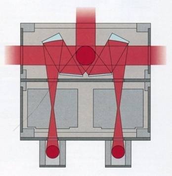
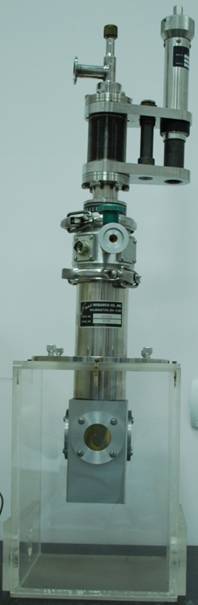
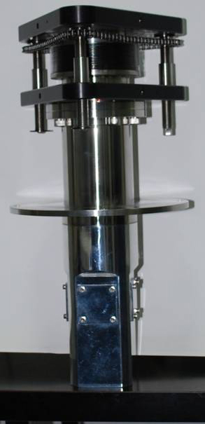
The configuration of the instrument is given in Fig. 2. It consists of
three sections: the upper one containing the source and the beamsplitter
compartment, the middle section with the beam switching compartment, the
sample compartment and the detector modules, and the lower part
containing the vacuum leads, the power supplies and the data processing
and control electronics. Depending of the moving mirror
travel, the resolution ranges from 0.06
to 0.0026 cm-1. The instrument has two focused output beams
in a sample compartment and three parallel output beams as shown in Fig.
2. In the present configuration our DA8 system operates within the 10 to
25 000 cm-1 range with a maximal resolution of 0.02 cm-1.
As sources we use a Hg lamp, a Globar (SiC) and a Quartz lamp. The
beamsplitters are a 25 μm mylar film (10-125 cm-1), a
Hypersplitter (40-700 cm-1) KBr (500-5000 cm-1)
and a Quartz (4000 – 25 000 cm-1). The following detectors
are used: DTGS (far IR), MCT (77K, mid infrared), InSb (77K, near
infrared) and Si photodiode (visible). The spectrometer is equipped with
two cryostats: a He flow Janis STDA 100 (LN2 and a LHe, 4-400
K) and an ARS DMX 20 closed-cycle low vibrational cryostat (4.5-300K)
with vibration amplitudes in the nanometer range. The system was
recently upgraded from far IR and MID IR to the NIR and VIS ranges,
namely, a NIR LN2 InSb detector (IPH5000L) for the range
1800-8500 cm-1, a visible Si (IPH5600L) detector for the
range 8500-50 000 cm-1 and a Quartz visible beamsplitter
(IMB2100L) for the range 4000-25 000 cm-1. For data
acquisition and processing, the DA 8 uses the latest working version of
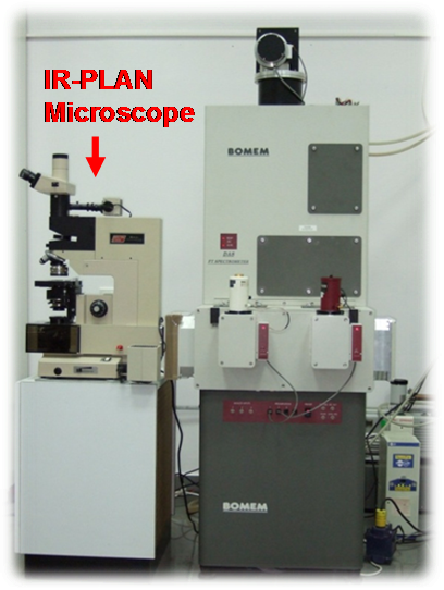
the Bomem GRAMS/AI 7.0 software. In addition, a suitable IR-PLAN advance
analytical microscope for measuring samples down to 7 µm in size is used
as shown in Fig. 1.
|
|
|
|
|
The DA8 system with 5 output ports. There are two focused output beams in a sample compartment and three parallel output beams. This model is appropriate for simultaneous installation of several experiments. |
Janis STDA 100 flow cryostat.
|
ARS DMX 20 low vibration closed-cycle cryostat. Cryostat chamber for Bomem DA 8 spectrometer. |
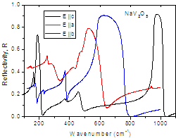
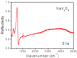
Some results
|
|
|
|
|
|
| Polarized FIR reflectivity spectra of NaV2O5 | E||a polarized FIR and MIR reflectivity spectrum of NaV2O5 |
IR-PLAN analytical microscope
IR-PLAN analytical microscope is a visible light microscope equipped with a high performance infrared sampling accessory designed for operation with FT-IR spectrometer. The microscope performance is directly related to the detection system because usually the analyzing samples are small so it is necessary to use MCT (mercury cadmium telluride) detector. IR-PLAN enables viewing of the exact sample area that will be analyzed and offers the best resolution available in FT-IR microscopy in order to obtain the highest possible quality spectra with the least stray light. IR-PLAN can be mounted, depending on the type of the spectrometer, in the primary compartment or in an external sample compartment or alongside of the spectrometer (Fig.1). IR-PLAN analytical microscope is equipped with a standard 1"x2" manual stage with a stage clip which provides the movement of the sample along the x, y and z-axis (focus adjustment). Usage of two circular, rotatable masking apertures above and below the sample reduces the stray light and other unwanted spectral contributions. The upper aperture is located in the infrared path between the infrared source and the sample whereas the lower aperture is located in the infrared path between the sample and the infrared detector. IR-PLAN analytical microscope can operate in transmission and reflectance mode.
Instrument specifications
Viewing optics:
Standard 15X Reflachromat IR/VIS objective of the Cassegrainian type
design for 150X viewing and 10X D-plan achromat glass objective for 100X
viewing and visual identification of the sample area with a larger field
of view.
Viewing attachments:
Binocular viewer has paired 10X eyepieces with crosshair measuring
reticle. In combination with the standard 15X objective, provides over
150X visual magnification.
Detection:
use of the spectrometer’s detector optics and detector or a dedicated
MCT detector.
Illumination:
High intensity reflected and transmitted light illumination with
variable light intensity and field and aperture stops.
Objective positioning:
4 position rotatable nosepiece.
Sample positioning:
Standard 2"x3"
travel rectangular rotatable
manual stage with stage clip.
Sample masking:
Two circular variable apertures for masking capable of being used
simultaneously to mask the sample, anywhere in the field of view.
Field of view& sampling area:
Nominal 1.3mm field of view with a 15x objective. Sampling area depend
on the detector, detector optics and the collecting objective being
used.
Purge: the spectrometer’s purge system can be used or own purge system
depending on the instrument and coupling.
Sampling mode:
Transmission or reflectance.
Microscope support:
Rolling work station/table.
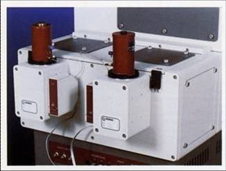
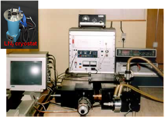
Polarising Fourier Transform Spectrometer SPECAC 40000 for FAR IR
The instrument is designed for operation in the spectral range between 3 and 250 cm-1 (90 GHz to 7.5 THz). It is configured as a polarisation interferometer using a single pair of wire grid polarisers acting as a beamsplitter and operates with either a phase or an amplitude modulation in a step-scan mode.
 |
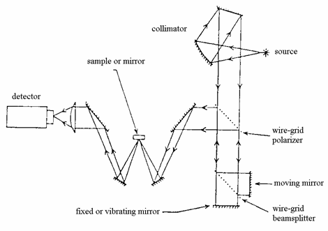 |
| SPECAC 40000 FAR IR spectrometer. Inset: LN2 cryostat | Configuration of SPECAC 40000 spectrometer for reflectance measurements |
The interferometer is based upon the polarising wire grid configuration developed by Martin and Puplett. The wire grids that are made from 5 mm diameter Tungsten wire spaced 12.5 (25) mm centre-to-centre. The first grid acts as a polariser to the collimated beam from the quartz-encapsulated mercury vapour arc lamp, producing two orthogonally plane polarised beams. The wires of the second grid are oriented at an angle of 45o relative to the first one, acting effectively as a beamsplitter.
The moving mirror has a mechanical path of 50 mm which in the case of a two-sided interferogram, gives the best resolution of 0.4 cm-1. The instrument is assembled with either a transmission or a reflection module for measurements on solid state specimens.
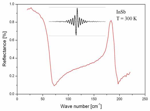
An advantage of this spectrometer is a phase modulation which has a better signal-to-noise ratio comparing to amplitude modulation. In the case of the phase modulation the fixed mirror vibrates with the frequencies between 10 and 20 Hz. As a consequence we have an asymmetric interferogram with the zero-leveled background as it is shown in the inset in figure down. For both modulations a lock-in amplifier must be used. The instrument works under the vacuum and is equipped with a LN2 cryostat.
|
|
|
FIR reflectivity spectrum of InSb at 300 K,
measured using Specac system |
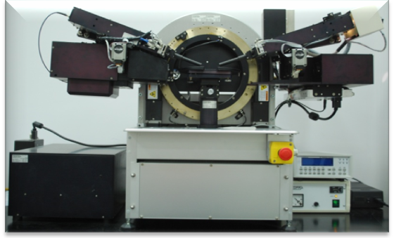
GES5E-IRSE Variable
Angle Spectroscopic Ellipsometer
The GES5E-IRSE Spectroscopic Ellipsometer is a combined system consisting of: DUV-Visible-NIR Spectroscopic ellipsometer (SE) and Fourier Transform Infra-Red Spectroscopic Ellipsometer (FTIR-SE). The polarizer and analyzer arms of both ellipsometers are mounted on a high resolution goniometric bench, made of double hollow crown. Both these crowns are driven by computer controlled stepper motors. The incidence angle can vary from 7 to 90° in DUV-Visible-NIR range, and from 20 to 90° in MIR range, with a theoretical resolution of 0.0005°.
|
|
|
|
The DUV-Visible-NIR Spectroscopic ellipsometer (SE) is working in a rotating polarizer configuration. It operates on the principle of mechanical modulation of the incidence light polarization by rotation of the polarizer at a constant angular frequency of 9 Hz. The analyzer remains in a fixed position, preparing the signal for the detector, insensitive to polarization. The precision of the ellipsometer for both weak and strong absorbing materials characterization is significantly enhanced with an automatically adjustable compensator. The light source is one 75 W Xe arc lamp, directly adapted to the polarizer arm. It emits a continuous spectrum of light, ranging from ultraviolet, trough visible to infrared (185-2000 nm). The light spot on the sample in parallel beam configuration is 1-10 mm2, depending on the aperture. There is also an additional miscrospot option for focusing the beam with a spot size of 365 x 270 μm, for an incidence angle of 75°. The light is introduced from the analyzer arm to the spectrometer using optical fiber making the light beam more stable. By combining the spectrometer with the photomultiplier tube (PMT) detector in UV-VIS range (190-900 nm), and InGaAs detector in NIR range (750-2000 nm), a high resolution spectrum is obtained by scanning the ellipsometric image at many discrete wavelengths. The spectrometer contains two dispersive elements (grating and prism) in collaboration with each other, separated by an intermediate fixed slit. The grating is blazed at optimum wavelength in order to obtain a maximum efficiency and the prism refracts the incoming wavelengths to act as a filter for higher order of diffraction produced by the grating.
The Fourier Transform Infra-Red Spectroscopic Ellipsometer (FTIR-SE) is a combination of a rotating analyzer configuration and a Fourier transform spectrometer. The basis of the FTIR spectrometer is a Michelson interferometer, which modulates each wavelength by a different frequency. The light leaving the Michelson interferometer enters the ellipsometer and successively passes the polarizer, the sample, the analyzer and finally hits the detector.
The light source for this IR ellipsometer is a silicon carbide (SiC) globar. Our system uses two different detectors: MCT in a range 580 cm-1 to 7000 cm-1, and DTGS in a range 385 cm-1 to 6500 cm-1. There is also an optional compensator in order to improve the accuracy of the measurements.
|
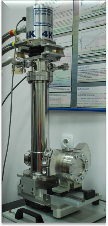
The ARS DX204 closed cycle low vibration
cryostat with additional DMX-20 interface allows for the low
temperature measurements up to 4.5 K. This cryostat is mounted
on a specially designed SOPRA stage and SOPRA chamber with three
possible angles of incidence (60°, 75°, 90°). |
 |
|
|
|
The spectroscopic ellipsometry can provide information about very thin layers, even down to a single atomic layer, or less. The measurements of the complex refractive index or dielectric function tensor give access to fundamental physical parameters that are related to a variety of sample properties, that include morphology, crystal quality, chemical composition, or electrical conductivity. It is commonly used to characterize single layer thin films or complex multilayer stacks ranging from a few parts of nanometers to several micrometers with an excellent accuracy.
 |
|
|
|
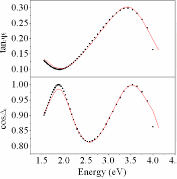
Measured ellipsometric parameters (circles) of thin films on a glass substrate and the best fit (full line) |
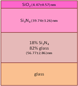
Best-fit model sketched |
Center
for Solid State and New Materials :: Facilities
:: Laboratory for the
Fourier transform Infrared (FTIR) spectroscopy and ellipsometry
::
print